How the Human Eye Works
Eyes. They help us so much in our daily lives. They help us to see and identify, which helps us to get stuff done. But there is more to the eye…. more than meets the eye . So welcome to this article that will tell you exactly how the human eye works. Stick around for the details involved and what occurs every time you spot something.

How The Eye Works
 How the Eye Is Designed
Learning about the structure and function of the eye in Biology has just increased my appreciation for the intricate parts of creation. Today, I thought I would share with you some information on how eyes work and the amazing things that go on when light is reflected into them.
The Ability to See and Capture
Your eye is your organ of sight. And it is a highly efficient, self-adjusting ‘camera’ that transmits impulses to the brain, where the object (focused on the eye’s retina) is interpreted as sight.
Did you know, that in sighted people vision actually supplies about 80-90% of the sensory information that reaches our brains? Incredible. All the colours, shapes and range of distances we can see would be difficult to perceive without the sense of sight.
So what eye structures and functions enable us to see? ….
The Structures and Functions of Your Eyes
Your eye essentially has three layers, the external, intermediate and internal.
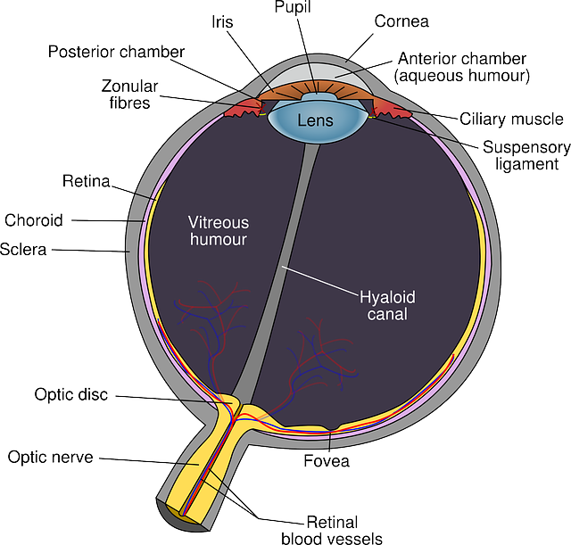
 Sketch of The Eye Structures
The Outer Eye
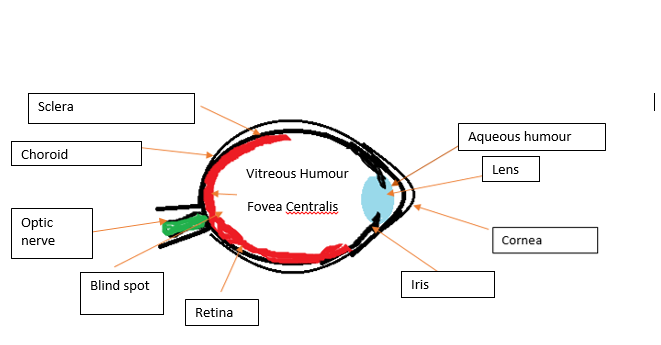
The external layer of the eye is a white tough layer, called the Sclera. This outermost layer protects and maintains the shape of the eye.
The Transparent part of the sclera – right at the front of the eye- is called the cornea. Light enters the eye through the cornea. The Cornea helps to refract or bend light through the pupil.
The Middle
The Choroid is the dark intermediate/middle layer of the eye.
The Choroid contains blood vessels that nourish the eye.
Also, the choroid absorbs stray light that enters your eye.
Iris
The Iris is the coloured part of the eye. Thin circular muscles of the Iris control the amount of light that enters the eye by changing the shape of the pupil.
Fun fact! Just like fingerprints, the pattern of colour in the Iris is unique to each person. That is why Iris scans can be used for personal identification.
The Pupil
The Pupil is the opening for light to enter the inner eye. The Iris adjusts the size of the pupil based on the light conditions – and this process is called adaptation.
You may have noticed that if you turn off the lights for a while, then put them on again, your pupils shrink. This is how your eye adapts to the changing light conditions. When the pupils shrink, it allows less light to enter your eyes, which makes it a bit more comfortable for you.
The Lens
The Lens sits behind the pupil and is attached to the ciliary muscles by suspensory ligaments. The Lens is a biconcave elastic structure which changes shape to help focus the image on the retina.
The Lens divides the eye into two chambers, the anterior chamber (in front of the lens) and the posterior chamber (behind the lens).
The Chambers
In the anterior chamber, a clear, watery fluid called the aqueous humour maintains the shape of the cornea. The aqueous humour also provides oxygen and nutrients for the surrounding cells, including the cells of the lens and cornea.
The posterior chamber is surrounded by the retina and contains a jelly-like substance called the vitreous humour. The vitreous humour helps to maintain the shape of the eyeball.
The Inner Eye
- The Retina is the internal layer of the eye. The Retina contains the photoreceptors that respond to light. Photoreceptors in the eye are rods and cone.
- Rods are sensitive to dim light, and involved in black and white and peripheral vision.
- On the other hand, Cones are sensitive to different wavelengths of light. Cones are involved in colour vision, bright light and detailed vision.
- The cones are packed most densely in the back of the eye in an area called the fovea centralis. So the fovea centralis contains a high concentration of cones, but no rods and it is the focal point of small detailed images.
- The rods and cones send sensory impulses to the brain via the optic nerve. The brain processes and integrates the information, and then perceives it as an image.
The Accommodation Reflex of the Lens
Because the Lens is flexible, it can change shape. This allows for finer focus when viewing objects, whether they are close or further away.
- For distant objects, the ciliary muscles relax and the lens becomes flat.
- For close objects, the ciliary muscles contract and the lens becomes round.
If you focus your vision on one thing for a long time (like reading a textbook up close for too long), the ongoing contraction of your ciliary muscle will likely cause muscle fatigue, which you will experience as eyestrain.
Vision Defects
Nearsightedness
- This is when a person can see close objects well, but not at a distance. The eyeball is elongated so that the image of a distant image is brought to focus in front of the retina, instead of on the photoreceptors.
- This is corrected by wearing concave lenses or by laser treatments – which removes cells from the cornea, making it more concave.
Farsightedness
- This is when a person can see distant objects well, but cannot focus on nearby objects because the eyeball is too short.
- The image of a close object is brought to focus behind the retina.
- This is corrected by wearing convex lenses which bend light rays at a sharper angle.
Astigmatism
This occurs when the cornea or lens is uneven. The image becomes fuzzy since light rays do not focus evenly. This is corrected by wearing an unevenly ground lens.
Glaucoma
- Glaucoma results from blockage of the ducts that drain the aqueous humour. When these ducts become plugged, pressure builds up in the eye.
- Increased pressure prevents blood flow to the retina. A lack of nutrients to the retina leads to partial or full blindness. Glaucoma can be relieved by surgery or medication.
Cataracts
As the Lens ages, its protein structure can start to degenerate, making it opaque and difficult for light to pass through. This condition can cause grey/white spots called cataracts on the lens. Cataracts is correctable using lasers or implants.
How Do You Get Depth Perception?
Depth perception is due to eye each forming an image from a slightly different angle. Some optic nerves cross over at the optic chiasma in the brain.
This means that the brain integrates and processes the various pieces of visual information, and images on each half of the brain are interpreted as a whole. The result is depth perception and three dimensional images. Pretty cool, eh?
The Human Eye is a work of art. But it is more than beautiful, as I hoped this article helped you to see!
Thanks for reading my mini guide on How the Human Eye Works.
Thanks for reading! If you liked this content, share with a friend:
Recent Articles
-
5 Best Media Training Courses to Prepare You for a Media Interview
Apr 10, 24 05:37 PM
 Preparing for an upcoming media interview? Here are the best online media training courses to equip you with the tools you need to succeed.
 -
5 Best Research Methods Courses and Certificates Online (2024)
Mar 27, 24 05:06 PM
 Here are the best research methods courses online that enable students and professionals to learn at their own pace and earn a certificate in research methods.
 -
The Fear of Crying | Why People are Scared to Cry, But Why You Should
Mar 27, 24 04:06 PM
 Are you scared to cry? Do you ever hold back your tears or hide them when you're in front of other people? Let's explore the fear of crying.